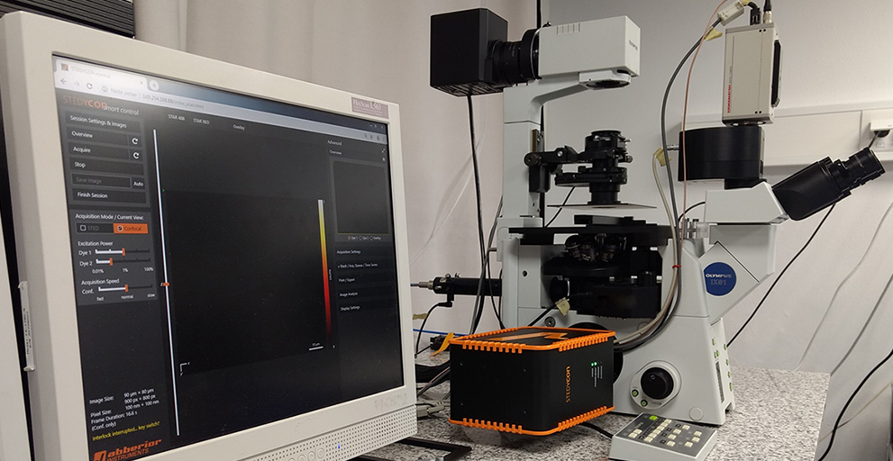
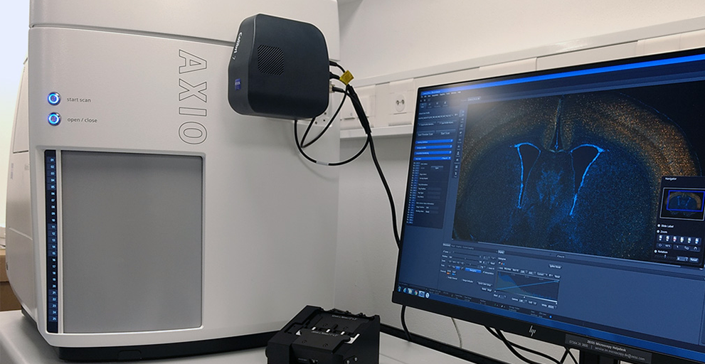
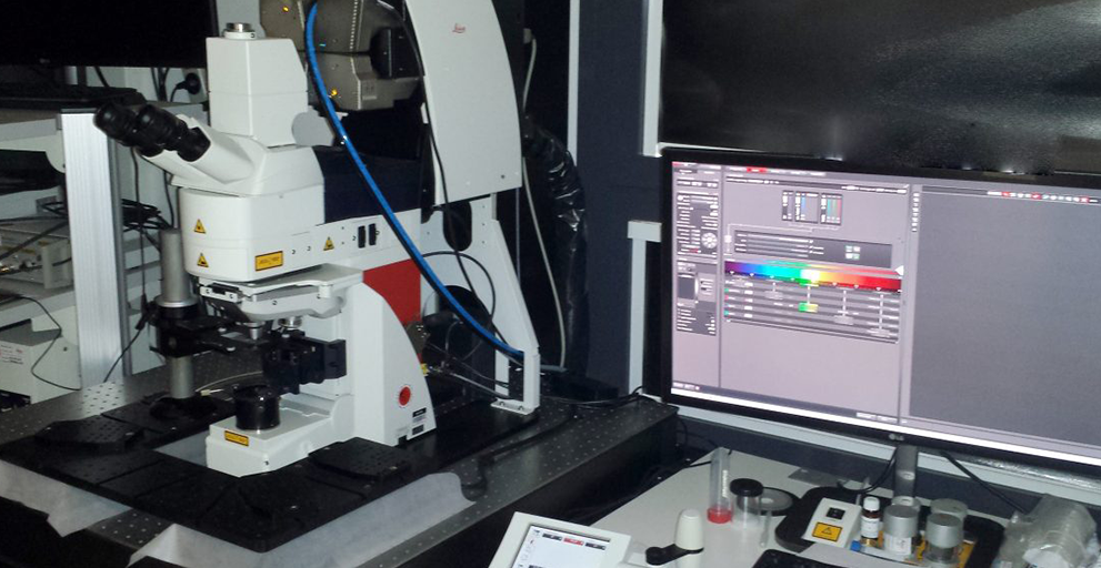
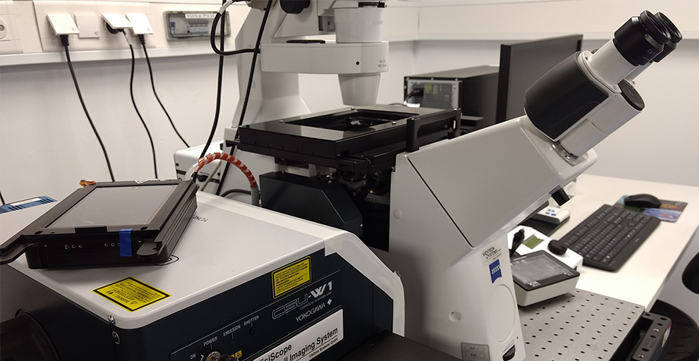
The Microscopy Core Facility provides access to several high-end microscopes at two locations at the Venusberg. At the BMZ 2 (Building 12) the instruments are specifically suited for imaging living cells while at the neurology (Building 82) the microscopes are oriented towards measuring (fixed) tissue sections and large organs. Furthermore, at the neurology we also offer electron microscopy and sample preparation for EM. For more details have a look at the electron microscopy page.
The table below gives you an overview of the different light microscopes installed at the MCF. The “search” field below can be used to filter the entries of the table so that you can identify microscopes, which provide the required features. E.g. you can find “confocal” or “super resolution” microscopes that are made for “fixed” or “live” imaging.
Feel free to contact us and we will discuss your projects with you to find the most appropriate imaging technology for your needs.
| Microscope | Type (general) | Type (specific) | Stand | Special features | Contact person | Location |
|---|---|---|---|---|---|---|
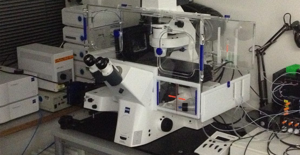
| Zeiss Observer.Z1 | Widefield | Inverted | Live cell imaging, dual cam | Gabor Horvath | BMZ 2 | |
| Olympus IX81 | Widefield | Inverted | Hannes Beckert | NER | ||
| Zeiss AxioScan.Z1 | Widefield | Slide scanner | Boxed (upright) | Fully automated, Robotics for 100 slides | Hannes Beckert | NER |
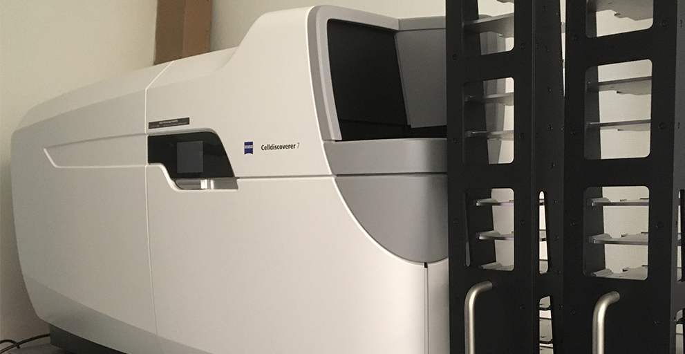
| Zeiss CellDiscoverer 7 | Widefield | High-content screening | Boxed (inverted) | Fully automated, Robotics for 24 plates | Gabor Horvath | BMZ 2 |
| Leica SP5 | Confocal | Point scanner | Inverted | Live cell imaging, FLIM, FCS | Gabor Horvath | Anatomy |
| Leica SP8 | Confocal | Point scanner | Upright | Cleared tissue imaging, FLIM, Long working distance objectives | Hannes Beckert | NER |
| Visitron VisiScope | Confocal | Spinning Disk | Inverted | Large tissue imaging, live cell imaging | Hannes Beckert | NER |
| Abberior STEDYCON | Confocal / Super-resolution | Point scanner / STED | Inverted | Hannes Beckert | NER | |
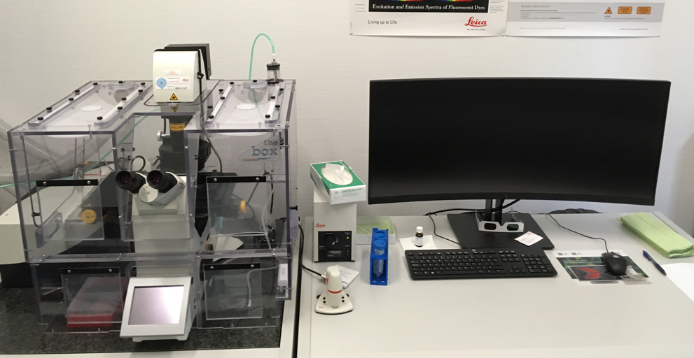
| Leica SP8 Lightning | Confocal / Super-resolution | Point scanner | Inverted | Live cell imaging, Deconvolution | Gabor Horvath | BMZ 2 |
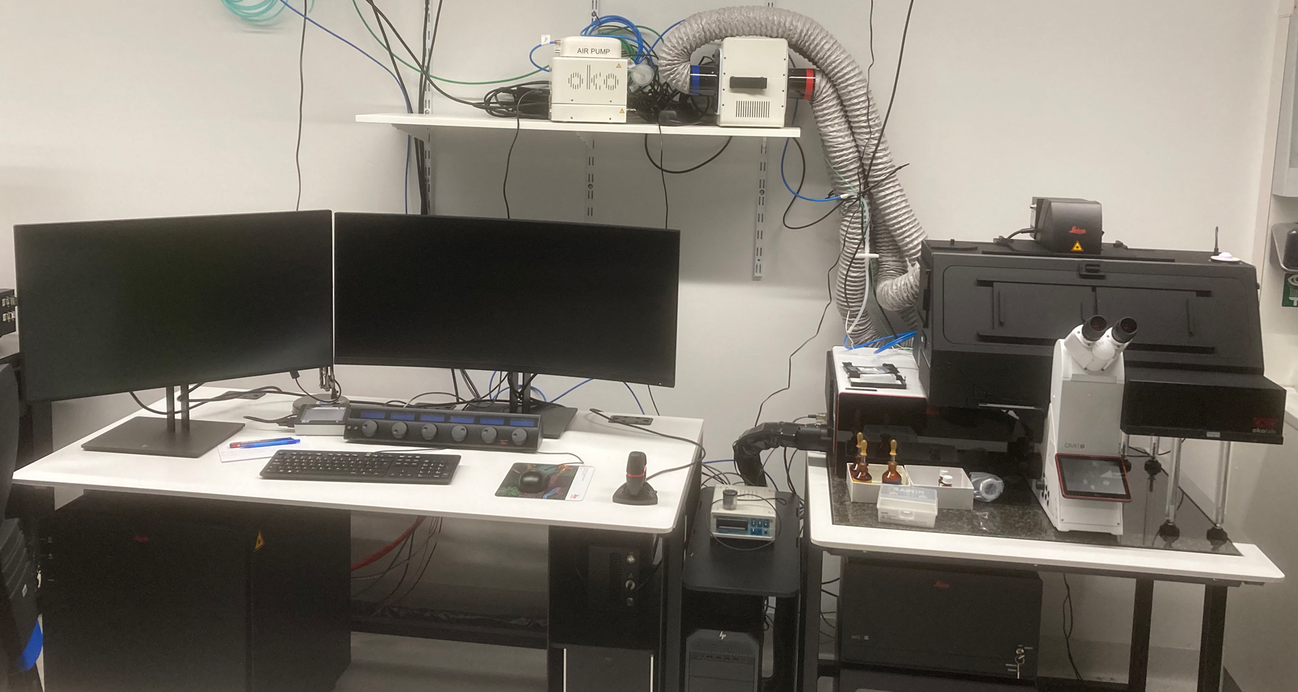
| Leica Stellaris 8 | Confocal / Super-resolution | Point scanner | Inverted | Live cell imaging, Deconvolution, FLIM | Gabor Horvath | BMZ 2 |
| LaVision Ultramicroscope II | Widefield | Lightsheet | Upright | Large, cleared specimen imaging | Gabor Horvath | BMZ 2 |
| Zeiss LSM980 Airyscan 2 | Confocal / Super-resolution | Point scanner | Inverted | Live cell imaging, Deconvolution, Spectral detection | Hannes Beckert | NER |
Location: BMZ 2 (Geb. 12)
The instruments at this site are specifically suited for imaging living cells. If you want to use one of the microscopes from that location, please contact Dr. Gabor Horvath.
Wide-field microscopes |
Confocal microscopes |
||
|---|---|---|---|
|
|
|
|
|
Location: Neurozentrum (Geb. 82)
The instruments at this site are oriented towards measuring tissue sections and large organs. If you want to use one of the microscopes from that location, please contact Dr. Hannes Beckert.
Wide-field microscopes |
Confocal microscopes |
|||
|---|---|---|---|---|
|
|
|
|
|
|
We also have a Zeiss Crossbeam 550 – an scanning electron microscope (SEM) with a focused ion beam (FIB). If you are interested in investigating the ultra structure of samples, please also get in contact with Dr. Hannes Beckert.

