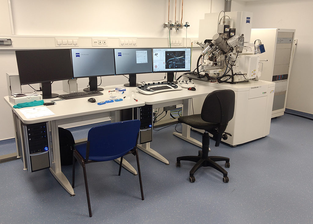
In the Microscopy Core Facility we also offer electron microscopy and sample preparation for EM. In most cases we will define a project and set up a price based on that project. So, do not hesitate to contact us to discuss your proposed projects. Most often the electron microscope will be operated by core personnel, however under special circumstances we also consider training people on the EM or sample preparation.
If you are interested in investigating the ultra structure of samples, please contact us at mcf_at_ukbonn.de
Sample Praparation and EM technologies
The MCF offers the following sample praparations and electron microscopy techniques:
- Embedding and heavy metal staining for conventional EM (e.g. EPON, Durcupan)
- Ultramicrotomy and collection of ultrathin sections
- Freeze substitution for correlated light and electron microscopy (e.g. in HM-20) [in development]
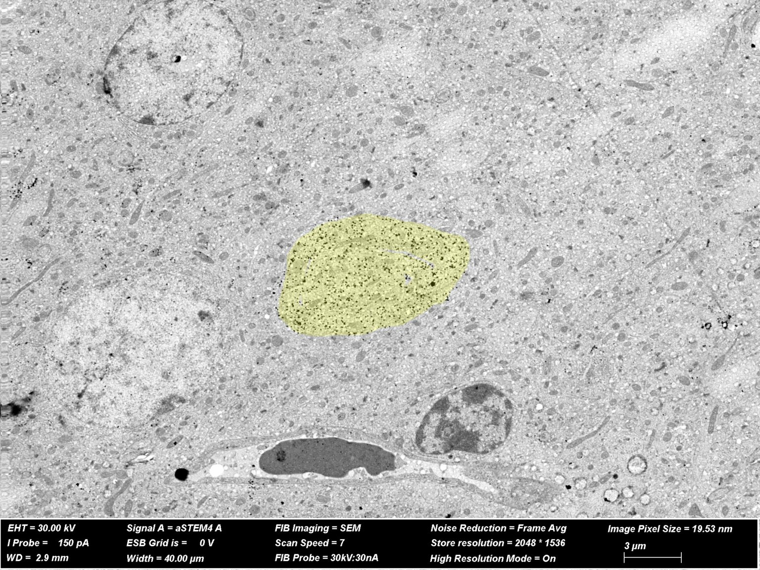
- Immunogold staining (pre- / post-embedding)
- Array tomography [in development]
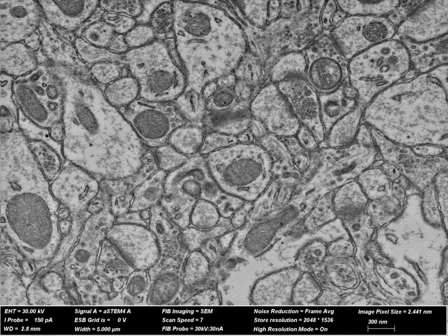
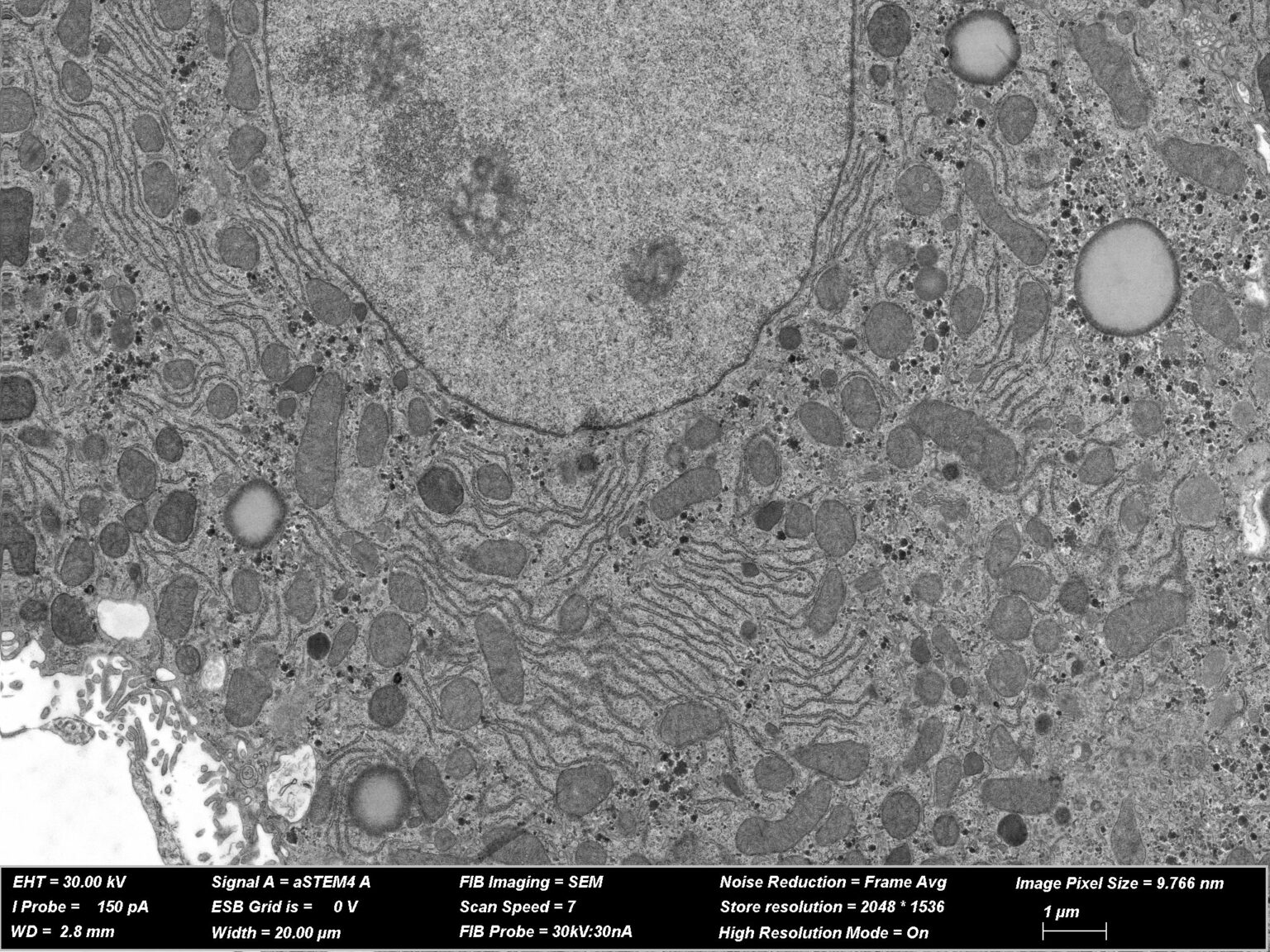
- 3D Nanotomography (Dual beam, FIB-SEM)
- Scanning transmission electron microscopy
Publications/Acknowledgment
Kim H, Melliti N, Breithausen E, Michel K, Colomer SF, Poguzhelskaya E, Nemcova P, Ewell L, Blaess S, Becker A, Pitsch J, Dietrich D, Schoch S., 2024. Paroxysmal dystonia results from the loss of RIM4 in Purkinje cells. Brain. 2024 Mar 13:awae081. https://doi.org/10.1093/brain/awae081
Paulussen, I., Beckert, H., Musial, T.F., Gschossmann, L.J., Wolf, J., Schmitt, M., Clasadonte, J., Mairet-Coello, G., Wolff, C., Schoch, S., Dietrich, D., 2023. SV2B defines a subpopulation of synaptic vesicles. Journal of Molecular Cell Biology mjad054. https://doi.org/10.1093/jmcb/mjad054
Kapadia, A., Theil, S., Opitz, S., Villacampa, N., Beckert, H., Schoch, S., Heneka, Michael.T., Kumar, S., Walter, J., 2023. Phosphorylation-state dependent intraneuronal sorting of Aβ differentially impairs autophagy and the endo-lysosomal system. Autophagy 1–22. https://doi.org/10.1080/15548627.2023.2252300
Próchnicki, T., Vasconcelos, M.B., Robinson, K.S., Mangan, M.S.J., De Graaf, D., Shkarina, K., Lovotti, M., Standke, L., Kaiser, R., Stahl, R., Duthie, F.G., Rothe, M., Antonova, K., Jenster, L.-M., Lau, Z.H., Rösing, S., Mirza, N., Gottschild, C., Wachten, D., Günther, C., Kufer, T.A., Schmidt, F.I., Zhong, F.L., Latz, E., 2023. Mitochondrial damage activates the NLRP10 inflammasome. Nat Immunol 24, 595–603. https://doi.org/10.1038/s41590-023-01451-y
Antoniou, A., Auderset, L., Kaurani, L., Sebastian, E., Zeng, Y., Allahham, M., Cases-Cunillera, S., Schoch, S., Gründemann, J., Fischer, A., Schneider, A., 2023. Neuronal extracellular vesicles and associated microRNAs induce circuit connectivity downstream BDNF. Cell Reports 42, 112063. https://doi.org/10.1016/j.celrep.2023.112063
Dietrich, A., Steffens, U., Gajdiss, M., Boschert, A.-L., Dröge, J.K., Szekat, C., Sass, P., Malik, I.T., Bornikoel, J., Reinke, L., Maček, B., Franz-Wachtel, M., Nieselt, K., Harbig, T., Scherlach, K., Brötz-Oesterhelt, H., Hertweck, C., Sahl, H.-G., Bierbaum, G., 2022. Cervimycin-Resistant Staphylococcus aureus Strains Display Vancomycin-Intermediate Resistant Phenotypes. Microbiol Spectr 10, e02567-22. https://doi.org/10.1128/spectrum.02567-22
Arévalo, L., Merges, G.E., Schneider, S., Oben, F.E., Neumann, I.S., Schorle, H., 2022. Loss of the cleaved-protamine 2 domain leads to incomplete histone-to-protamine exchange and infertility in mice. PLoS Genet 18, e1010272. https://doi.org/10.1371/journal.pgen.1010272
Merges, G.E., Meier, J., Schneider, S., Kruse, A., Fröbius, A.C., Kirfel, G., Steger, K., Arévalo, L., Schorle, H., 2022. Loss of Prm1 leads to defective chromatin protamination, impaired PRM2 processing, reduced sperm motility and subfertility in male mice. Development 149, dev200330. https://doi.org/10.1242/dev.200330
Müller, J.A., Betzin, J., Santos-Tejedor, J., Mayer, A., Oprişoreanu, A.-M., Engholm-Keller, K., Paulußen, I., Gulakova, P., McGovern, T.D., Gschossman, L.J., Schönhense, E., Wark, J.R., Lamprecht, A., Becker, A.J., Waardenberg, A.J., Graham, M.E., Dietrich, D., Schoch, S., 2022. A presynaptic phosphosignaling hub for lasting homeostatic plasticity. Cell Reports 39, 110696. https://doi.org/10.1016/j.celrep.2022.110696
Scheiblich, H., Dansokho, C., Mercan, D., Schmidt, S.V., Bousset, L., Wischhof, L., Eikens, F., Odainic, A., Spitzer, J., Griep, A., Schwartz, S., Bano, D., Latz, E., Melki, R., Heneka, M.T., 2021. Microglia jointly degrade fibrillar alpha-synuclein cargo by distribution through tunneling nanotubes. Cell 184, 5089-5106.e21. https://doi.org/10.1016/j.cell.2021.09.007
Zhang, L., Haddouti, E.-M., Beckert, H., Biehl, R., Pariyar, S., Rüwald, J.M., Li, X., Jaenisch, M., Burger, C., Wirtz, D.C., Kabir, K., Schildberg, F.A., 2020. Investigation of Cytotoxicity, Oxidative Stress, and Inflammatory Responses of Tantalum Nanoparticles in THP-1-Derived Macrophages. Mediators of Inflammation 2020, 1–14. https://doi.org/10.1155/2020/3824593