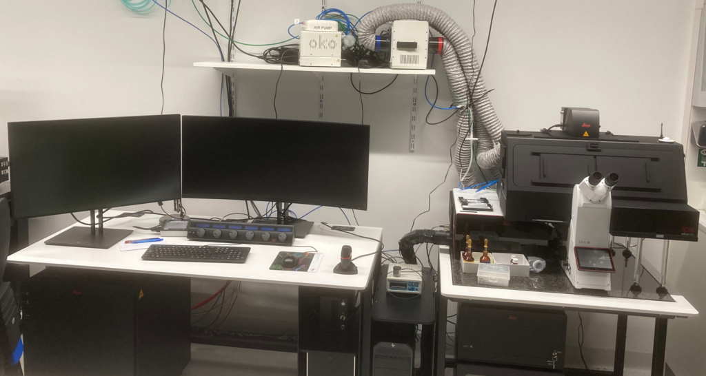
Location: BMZ 2, 2OG.303
Technical information:
Objectives
| Magnification | Type | NA | Immersion |
|---|---|---|---|
| 10X | HC PL APO CS2 | 0.40 | dry |
| 20X | HC PL APO CS2 | 0.75 corr | any |
| 40X | HC PL APO CS2 | 1.25 motcorr | glycerol |
| 63X | HC PL APO CS2 | 1.20 corr | water |
| 63X | HC PL APO CS2 | 1.40 | oil |
Lasers
405 nm
White Light Laser (440 – 790 nm)
Accessories
5 x Power HyD detectors (2 x HyD S, 2 x HyD X, and 1 x HyD R)
Resonance scanner (8 kHz) for fast live imaging with minimal photo-toxicity
dark environmental chamber
CO2-incubation
automated water-immersion pump
Software
LAS X v4.5.0
Wizards: FRAP, FRET, Lightning, Navigator
ImageCompass
Simplified user interface, even for complex experiments. Set up a multi-color experiment with one click per fluorophore. Maximum signal yield through automatically selected optimal recording settings. Multi-dimensional capture, full control of motorized hardware. GUI optimized for dark rooms and image processing.
Lightning Deconvolution Software
The Lightning module automatically detects the finest structures and details. The key is an adaptive process for extraction of hidden information in the image. Unlike traditional deconvolution techniques, that use a global set of parameters for the full image, Lightning calculates an appropriate set of parameters for each voxel to uncover every detail with the highest fidelity.
The Lightning detection concept extends the confocal microscope by simultaneous multi-color super-resolution imaging at the full original speed.
TauSense
Instant access to information about the average photon arrival time and intensity (TauContrast). Differentiation between desired and undesired fluorescence signal (TauGating, GateScan). Separation of fluorophores based on their fluorescence lifetime (TauSeparation, TauScan).
FALCON – Fast Lifetime Image Contrast Software
STELLARIS 8 FALCON is a FLIM solution for fast acquisition, processing and analysis of FLIM images. The software is fully integrated in LAS X and allows FLIM measurements with any measurement mode (time series, 3D measurements, lambda scan, can be combined with Mosaic measurements (LAS X Navigator)). Several detectors can be combined to form a FLIM detector to speed up acquisition. The so-called fast FLIM image (mean time of arrival) and a photon pulse histogram are displayed in real time. LAS X FLIM contains an evaluation mode specialized for FLIM-FRET. Here, in particular, images of the apparent FRET efficiency, the FLIM FRET efficiency and binding ability. FALCON includes Phasor FLIM Analysis, which enables acquisition and analysis of FLIM images using the Phasor method. FLIM Phasors provide a 2D graphical view to distinguish and separate different lifetime types within an image.
Funding
Please acknowledge the use of the instrument in publications the following way:
“We would like to thank the Microscopy Core Facility of the Medical Faculty at the University of Bonn for providing support and instrumentation funded by the Bundesministerium för Building und Forschung (BMBF, Federal Ministry of Education and Research) – ACCENT: Förderung von Advanced Clinician Scientist im Bereich Immunopathogenese und Organdysfunktion, Gehirn und Neurodegeneration – Förderkennzeichen: 01EO2107.”