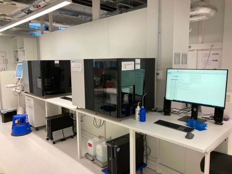
Many of you will already know that the FCCF could expand its equipment park considerably this year.
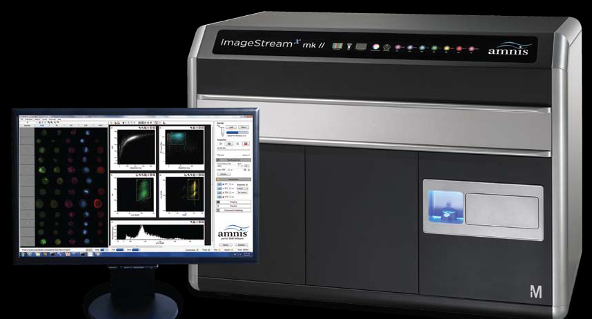
By now, all instruments are delivered, but not all of them are ready to use, due to different reasons. The first instrument we wanted to introduce to you, is the ImageStream®X MarkII by Amnis®.
This cytometer offers our users a technology wich combines the best out of two worlds:
Imaging Cytometry
Imaging flow cytometers combine the speed and sample size of flow cytometry with the resolution and sensitivity of microscopy in a single instrument platform unlike any other available for cell analysis. With up to 12 channels for each cell in a population, microscopic images provide qualitative and quantitative image data of every event acquired in flow.
The instrument is now ready to use and we are looking forward to discuss with you your experiments you want to perform on this platform.
Since this technique is new in our hands, we will need your projects to gain more and more expertise. In the next months we plan to closely accompany the users of the device during the acquisition as well as during the analysis of the data afterwards.
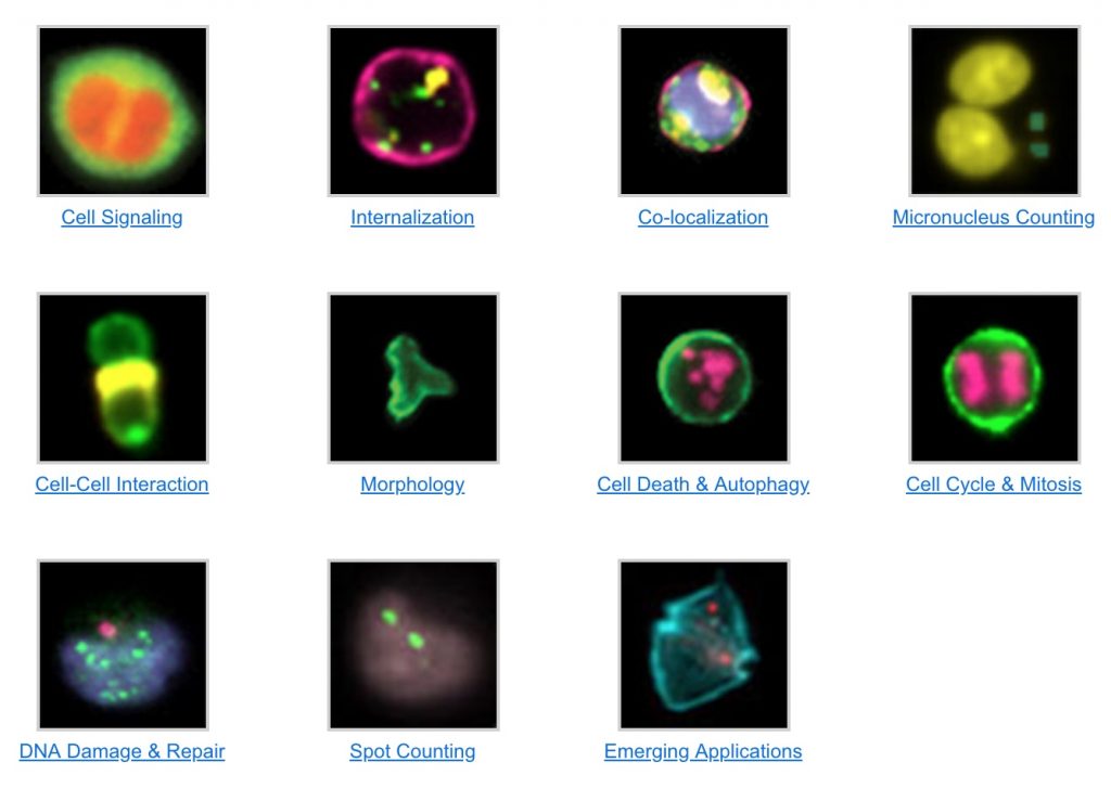
Follow the link to the ImageStream website, there you can find detailed informations about possible applications.
Our Instrument is fully equipt with 7 lasers:
A high power 488nm laser for enhanced detection of fluorescent dyes excited at that wavelength and six additional lasers, including 375, 405, 561, 592, and 642 nm for maximal experimental flexibility.
Up to 12 images of every cell:
The 12 Image Channel option significantly expands the experimental flexibility and analytical potential of the ImageStream®X Mark II Imaging Flow Cytometer by doubling the number of images per cell from six to twelve. This option is comprised of a second image detection system, including a filter wheel, inline filters, spectral decomposition assembly, and proprietary CCD camera.
60X / 40X / 20X Magnification:
The MultiMag option provides 20X and 60X objective lenses in addition to the standard 40X objective for greater experimental flexibility and improved resolution. The 60X objective increases magnification for small objects such as yeast and bacteria and offers greater detail with mammalian and plant cells. The 20X objective provides an increased field of view for the imaging of large cells, such as epithelial cells.
Extended Depth of Field:
A unique optical system upgrade for the ImageStream®X cytometer keeps the depth of cell in focus without loss of fluorescence sensitivity. Now you can image FISH spots or other sub-cellular features no matter where they lie in the cell.
Image More of the Cell in Focus
The extended depth of field (EDF™) option for Amnis® imaging flow cytometry system incorporates Wavefront Coding™ technology from CDM Optics, which is a combination of specialized optics and unique image processing algorithms, to project more structures within the cell into one crisp plane of focus.
AutoSampler for Multiwell Plates:
After the first experiments we witnessed, that imaging cytometry in fact is way more time consuming than conventional cytometry, so we decided to purchase the AutoSampler option for the ImageStream® to enhance the productivity with unattended sample loading from mulitwell plates.
IDEAS®Software for data analysis:
Of course the analysis of imaging data can be very challenging. To support scientists in this task, we have also a campus license of the IDEAS® Software. It offers powerful, familiar tools for creating histograms and scatter plots. Click on any dot in a scatter plot to see the corresponding cell’s image. Numerical scoring of parameters such as size, shape, texture, colocalization and intensity allow quantification of visual data.
If we’ve aroused your interest, just contact us for a project discussion.